Study with Iris · Neuro · Clinical reasoning
A visual pattern-recognition tool for comparing the two links in voluntary motor control and the findings that usually travel with each pattern.
High-yield: group the cluster, trace the pathway, then check context.
Learning purpose
After reviewing this guide, you should be able to…
Why this matters
Weakness is not one pattern.
Weakness alone does not localize the pattern. Compare the finding cluster without turning a concise study tool into a diagnosis.
UMN
↑ tone · ↑ reflexes
descending-control cue
acute timing · mixed findings · context
LMN
↓ tone · ↓ reflexes
final-pathway cue
Fast first move
Weakness is the start, not the localization.
WEAKNESS
⌁ muscle cue
Start here.
COMPARE
⌁ reflex cue
tone · reflexes · plantar · bulk
LOCALIZE
⌁ pathway cue
descending control or final pathway?
PAUSE
⌁ timing cue
acute course · mixed findings
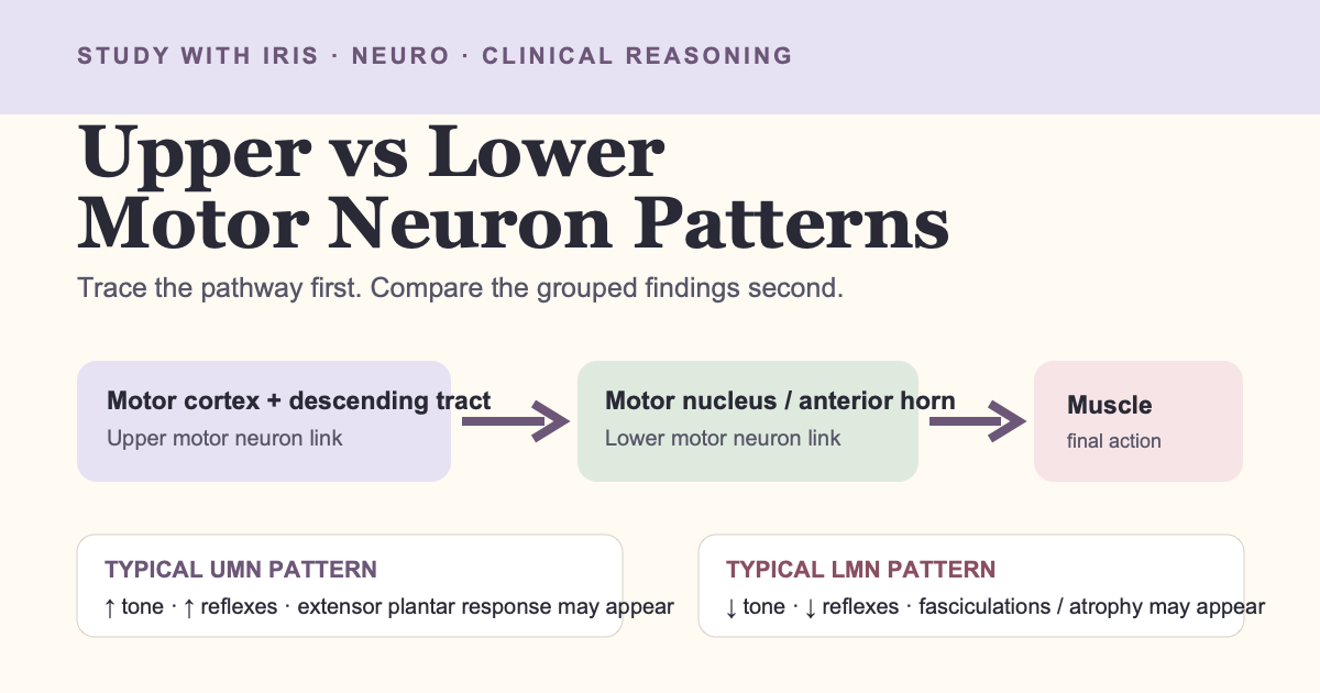
Teaching visual 1 · anatomy + localization
See the pathway. Find the breakpoint first.
typical UMN-pattern localization
typical LMN-pattern localization
descending control
→
motor nucleus / anterior horn
→
final common pathway
Teaching visual 2 · comparison + clinical reasoning
See the pattern in 10 seconds.
Typical UMN pattern
TONE + REFLEXES
increased / hyperactive
PLANTAR
extensor response may support corticospinal dysfunction
BULK + TWITCH
less early atrophy · fasciculations not typical
Typical LMN pattern
TONE + REFLEXES
decreased / diminished
PLANTAR
extensor response is not typical
BULK + TWITCH
atrophy can develop · fasciculations can occur
Swipe the table sideways to see all columns.
| Finding | Typical UMN pattern | Typical LMN pattern |
|---|---|---|
| Muscle tone | Increased tone or spasticity | Decreased or absent tone |
| Deep tendon reflexes | Hyperactive | Diminished or absent |
| Plantar response | Extensor response can support corticospinal dysfunction | Not the typical LMN-pattern finding |
| Atrophy | Not typical early; disuse-related atrophy can occur over time | Can become prominent over time |
| Fasciculations | Not typical | Can be present |
↑ or ↓?
↑ or ↓?
supporting cue
add context
Look closer
Two study traps worth correcting
“UMN always means stiff immediately.”
Acute severe central lesions can initially produce flaccid paresis, reduced tone, and reduced reflexes before an UMN pattern evolves. Do not ignore timing.
“LMN signs diagnose the cause.”
An LMN-like pattern supports localization; it does not establish one disease or replace a complete neurologic examination.
Original concept practice
Do the reasoning: Pattern → Why → Pause
Card A
Increased tone, brisk reflexes, and an extensor plantar response are grouped in a course vignette.
WHAT DO YOU NOTICE? → decide before peeking
Card B
Reduced tone, reduced reflexes, visible fasciculations, and progressive atrophy are grouped in a course vignette.
WHAT DO YOU NOTICE? → decide before peeking
Card C
New severe weakness is paired with low tone and low reflexes soon after an acute central event in a course vignette.
WHAT DO YOU NOTICE? → decide before peeking
Your turn · write before checking
Retrieve → Check
Try first. Then uncover the check.
Cover the answer row with a hand or note. Retrieve before you read.
RETRIEVE 1
Which two observations are most useful to compare first?
RETRIEVE 2
Where does the lower motor neuron begin on this simplified map?
RETRIEVE 3
What should make you pause before applying a memorized rule?
Check your reasoning
Tone and deep tendon reflexes are efficient opening observations; then add plantar response, atrophy, and fasciculations.
In this simplified map, the LMN begins at a cranial motor nucleus or anterior horn cell and continues through the peripheral pathway.
Acute timing, mixed or incomplete findings, and full context can make a memorized binary rule unreliable.
Case answer key
Why: increased tone, brisk reflexes, and extensor plantar response form a descending-control cluster.
Why: reduced tone/reflexes with fasciculations and atrophy fit a final-common-pathway cluster.
Why: acute central injury may initially look flaccid and hyporeflexic.
Sources
Verified references used for this guide
- Merck Manual Professional: Distinguishing Upper From Lower Motor Neuron Lesions
- Merck Manual Professional: Weakness
- NCBI Bookshelf: Neuroanatomy, Motor Neuron
Reviewed October 2026 · Visual Learning Revision 2.1
← Back to Study with Iris · Cranial Nerves Study Guide · ABG Pattern Recognition
Keep exploring
Helpful Iris resources for this topic
Free Scholarship Tool
Get the PTA to DPT Scholarship Tracker
Track deadlines, award amounts, essays, recommendations, submission dates, notes, and renewals without letting scholarship season become scattered.Get the free trackerShare This Article
Found this helpful?
Share it with another student, clinician, traveler, or future DPT who might need it.