DPT Study Guides
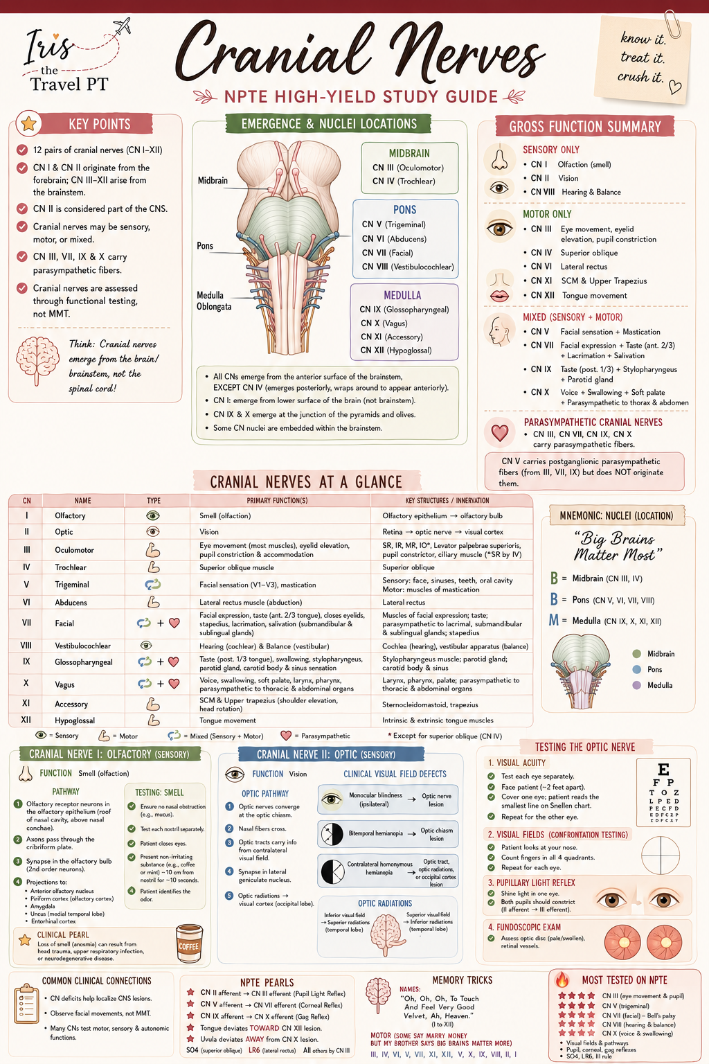
Cranial Nerves Study Guide
A visual cranial-nerve anatomy and clinical-reasoning guide for PT, PTA, and DPT learners to sort function, map broad regions, and use a focused examination framework with context.
Study with Iris · Neuro anatomy + clinical reasoning

Cranial nerves: organize first, then test with purpose.
A visual, first-pass framework for PT, PTA, and DPT learners. Use it to make the 12 nerves easier to sort, retrieve, and connect to a focused examination—not to diagnose a person from one finding.
After reviewing this guide, you should be able to…
Why this matters: Cranial-nerve questions become less like twelve disconnected facts when function, region, and a purposeful observation travel together.
See the route in 30 seconds.
Forebrain → brainstem map
Olfaction · vision
Eye movement group begins here
Face, eye abduction, facial movement, hearing / balance
Pharynx / voice, neck, tongue
What to remember
I–II: unlike the remaining nerves, they are associated with the forebrain rather than a brainstem exit.
III–XII: learn an introductory region map first, then add nuclei, course, and exceptions in your course materials.
Fast cue: 3–4 midbrain · 5–8 pons region · 9–12 medulla.
PAUSE / CONTEXT: This is a simplified learning map, not a complete nuclei or lesion map. Multiple cranial-nerve deficits, associated central signs, timing, and the actual examination all change interpretation.
Sort before you memorize details.
Sensory only
I · II · VIII
Smell · vision · hearing / balance
Motor only
III · IV · VI · XI · XII
Eye movement · neck / shoulder · tongue
Mixed
V · VII · IX · X
Face / jaw · facial movement · pharynx / voice
Traditional sensory/motor/mixed categories are a helpful first sort. They do not replace learning the individual sensory, motor, and autonomic components of each nerve.
Swipe the table sideways to see all columns.
| Group | Core function cue | Focused observation to connect | Do not overcall |
|---|---|---|---|
| I · II | Smell · vision | Smell history / visual function in the appropriate exam context | One observed change does not identify its cause. |
| III · IV · VI | Eye movement group | Extraocular movements and pupil-related findings are interpreted together | Do not assign a single ocular finding to one lesion without the full exam. |
| V · VII | Facial sensation / mastication · facial movement | Compare face sensation or movement thoughtfully and with symmetry in view | Facial pattern recognition is not a diagnosis. |
| VIII | Hearing / balance | Connect hearing or vestibular observations to the exam question | Balance symptoms have many possible contributors. |
| IX · X | Pharynx / palate / voice | Observe relevant voice, palate, and swallow-related features within supervision and setting | Do not turn a study guide into a swallow or airway decision tool. |
| XI · XII | Neck / shoulder · tongue | Connect shoulder elevation / head turn or tongue movement to the focused exam | Interpret with the rest of the neurologic examination. |
The table is fully visible in this printable version.
Apply a calm four-step study sequence.
Pattern card A
A prompt asks about lateral eye movement.
Start with: the III–IV–VI eye-movement group; then retrieve that CN VI supplies lateral rectus.
Pattern card B
A prompt pairs facial sensation with mastication.
Start with: CN V, a mixed nerve; then add its three facial sensory divisions in your course review.
Pattern card C
A prompt pairs shoulder elevation with head turning.
Start with: CN XI, a motor nerve; connect it to sternocleidomastoid and trapezius.
Retrieve first. Then check your reasoning.
1. Which three cranial nerves are traditionally classified as sensory only?
2. What is the fastest broad-region sequence for III–XII?
3. A learner identifies a facial movement asymmetry. What is the safe next thought?
Print answer key
1. I, II, and VIII. These are sensory-only in the traditional first-pass classification: smell, vision, and hearing / balance.
2. III–IV midbrain; V–VIII pons or pontomedullary region; IX–XII medulla. This is a broad organizing map, not a complete nuclei or course map.
3. CN VII is a relevant group, then pause. Interpret any facial finding with the complete examination, timing, associated signs, supervision pathway, and clinical context; a pattern does not establish a diagnosis.
IF YOU MISSED THIS: revisit the fast pattern board for function category, the route map for broad region, and the four-step sequence for the pause/context cue.
Sources and scope
- Sonne J, Omole AE, Lopez-Ojeda W. Neuroanatomy, Cranial Nerve. StatPearls / NCBI Bookshelf. Updated January 24, 2025. Supports the traditional sensory, motor, and mixed classification and core functions.
- Casselman JW, Krainik A, Macdonald I. Evaluation of Patients with Cranial Nerve Disorders. Diseases of the Brain, Head and Neck, Spine 2024–2027. NCBI Bookshelf. Supports the simplified forebrain / brainstem regional associations and the caution that lesion interpretation requires an adapted clinical evaluation.
- Yaddanapudi S. How To Do the Cranial Nerve Examination. Merck Manual Professional Edition. Supports the examination-context framing.
This is an educational anatomy and clinical-reasoning resource for students. It does not replace course instruction, clinical supervision, an examination protocol, medical advice, or clinical judgment. If a real person has new or concerning neurologic symptoms, follow the setting’s escalation pathway rather than using a study guide to interpret or manage them.
← Back to Study with Iris · Upper vs Lower Motor Neuron Patterns · Muscles of Facial Expression
DPT Study Guides
Keep exploring
Keep exploring
Helpful Iris resources for this topic
Share This Article
Found this helpful?
Share it with another student, clinician, traveler, or future DPT who might need it.