DPT Study Guides
Muscles of Facial Expression Study Guide
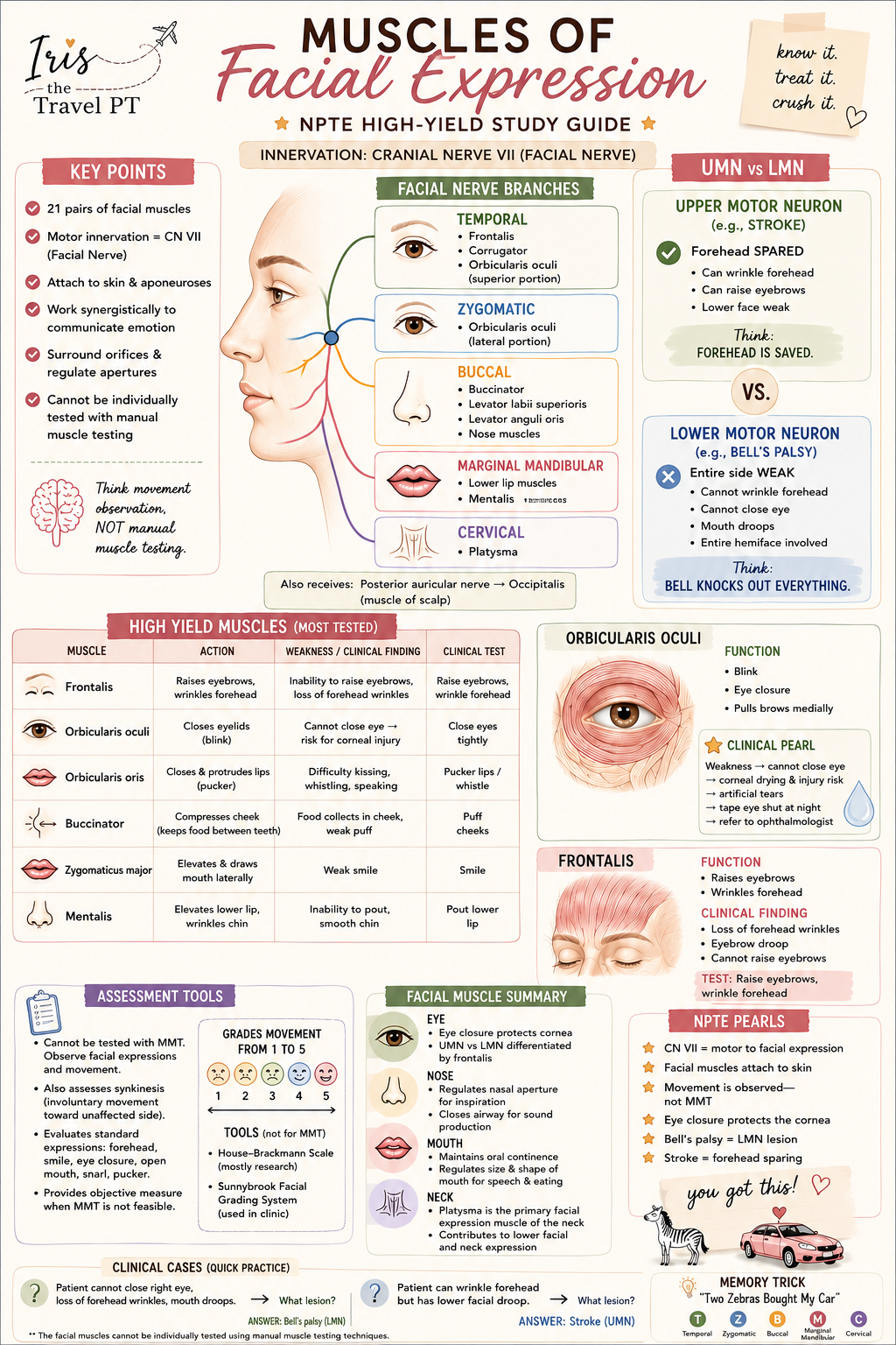
A visual neuroanatomy and clinical-reasoning study guide for connecting facial movements, broad facial nerve territories, and pattern-based examination thinking.
Study with Iris – neuroanatomy + clinical reasoning

Facial movement: name the task, then locate the pattern.
Use this guide to connect a facial movement to a muscle group, a facial-nerve branch, and the examination pattern that deserves context. It is a study framework, not a diagnostic shortcut.
Start with the movement.
Use broad territories before details
Upper face: brow elevation and eye closure help organize forehead and periocular movement.
Midface: cheek and smile tasks connect with zygomatic and buccal branch territories.
Lower face: lower-lip depression and related movement provide a separate observation point.
The named branches and individual muscles have overlap and variation. For studying, use this map as an organizing scaffold, then add course-specific anatomy.
Compare the pathway, not one isolated sign.
What the comparison means
A central pathway pattern may affect voluntary lower facial movement more visibly than forehead movement because upper facial motor control has bilateral cortical contribution in the classic teaching model.
A peripheral facial-nerve pattern can involve upper and lower facial movement on the affected side. Those patterns still require context; this guide does not establish cause or urgency.
Study cue: “Upper face has bilateral cortical input; lower face is more crossed.” Then ask what else is present.
Make the exam observation useful.
Swipe the table sideways to see all columns.
| Movement cue | Muscle group to retrieve | Broad branch territory | Reasoning reminder |
|---|---|---|---|
| Raise brows | Frontalis | Temporal | Compare both sides; do not use one task alone to localize. |
| Close eyes tightly | Orbicularis oculi | Temporal / zygomatic | Observe closure quality and the rest of the facial pattern. |
| Smile / show teeth | Zygomaticus group, levator group | Zygomatic / buccal | Lower-face asymmetry needs the whole neurologic context. |
| Depress lower lip | Depressor labii / anguli groups | Marginal mandibular | Branch territories are a learning aid, not an isolated diagnostic test. |
Test the framework before you reread.
A learner can raise both eyebrows but has new lower-face asymmetry. What is the high-yield next thought?
Which observation best keeps a facial-movement exam structured?
What does the branch map help you remember?
Sources and study note
- Blumenfeld H. Neuroanatomy Through Clinical Cases. 3rd ed. Sinauer Associates; 2021.
- National Institute of Neurological Disorders and Stroke. Facial Paralysis Information Page. Accessed October 2026.
Educational resource for learners and clinicians. In real care, follow clinical supervision, local protocols, and urgent-assessment pathways when indicated.
Continue learning
DPT Study Guides
Keep exploring
Keep exploring
Helpful Iris resources for this topic
Share This Article
Found this helpful?
Share it with another student, clinician, traveler, or future DPT who might need it.